Updated: Feburary 10, 2026
CSF is a clear, colorless fluid that appears similar to water, but don’t let its simple appearance fool you. This plasma-like substance has a carefully regulated composition that remains remarkably constant under normal conditions. The specific gravity ranges from 1.003 to 1.008, making it slightly denser than water, while the pH stays between 7.31 and 7.40, maintaining a slightly alkaline environment crucial for proper brain function.
Under normal circumstances, CSF contains no bilirubin and extremely low cholesterol levels. This absence is significant because the presence of these substances immediately signals abnormal conditions requiring medical attention.
Healthy CSF contains very few cells. The normal white blood cell count should be less than 5 cells per microliter, with the majority being lymphocytes and monocytes. Red blood cells should be completely absent. When these numbers change, they provide crucial diagnostic clues.
The appearance of white blood cells, particularly neutrophils, often indicates infection or inflammation. In bacterial meningitis, for instance, white blood cell counts frequently exceed 1,000 per microliter, with polymorphonuclear leukocytes predominating. Viral infections typically produce a milder elevation with lymphocyte predominance.
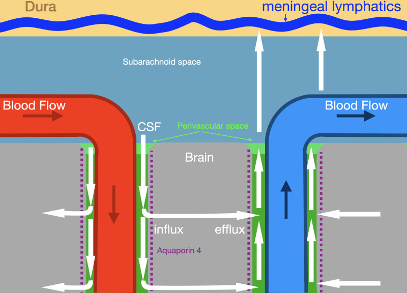
The illustration shows glymphatic fluid flow (white arrows) along periarterial and perivenous pathways. Labeled structures include the dura, meningeal lymphatics, subarachnoid space, brain parenchyma, arterial and venous circulation, and aquaporin 4 channels mediating influx and efflux. Contributed by Konstantinos Margetis MD, PhD
Concentration of sodium, potassium and urea are similar to those found in the plasma.
Sodium and chloride are the most abundant ions.
The normal adult CSF volume is about 130 ml.
Glucose serves as the brain’s primary fuel source measuring from 60-80% of blood glucose levels.
Beyond infections and hemorrhage, CSF composition helps diagnose various neurological disorders. In multiple sclerosis, the presence of oligoclonal bands, specific proteins that appear only in CSF and not in blood, now substitutes for traditional disease progression criteria in patients younger than 50 years with typical clinical presentations.
Guillain-Barré syndrome produces a characteristic pattern called albuminocytologic dissociation, where protein levels rise significantly while cell counts remain normal. This finding appears in 50% of patients during the first week, increasing to 75% by three weeks.
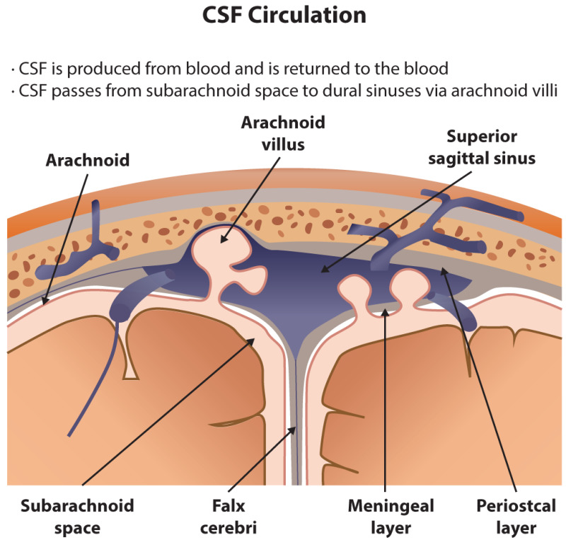
Understanding normal CSF composition provides the foundation for recognizing disease. Your body produces approximately 500 mL of CSF daily while maintaining a constant volume of 125 to 150 mL, completely replacing the fluid every 7.5 hours. This constant turnover ensures that changes in composition rapidly reflect ongoing disease processes.
The blood-brain barrier tightly regulates what enters CSF, maintaining its unique composition. When disease disrupts this barrier, plasma proteins leak into CSF, electrolyte ratios shift, and cells that normally stay in blood vessels migrate into the fluid. Each change tells part of the diagnostic story.
The collection technique also matters. Opening pressure measured during lumbar puncture provides valuable information about intracranial conditions. Multiple tubes collected in sequence help distinguish true bleeding from traumatic taps. Proper handling and rapid analysis preserve cellular elements that deteriorate if samples sit too long.
There are different ways to get a sample of CSF. Lumbar puncture (spinal tap) is the most common method.
To have the test:
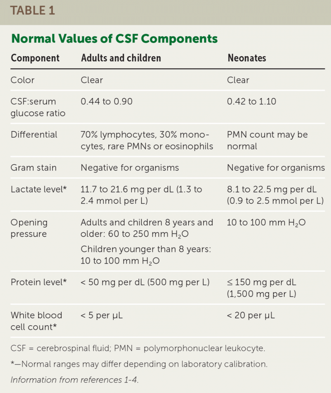
Normal values typically range as follows:
Normal value ranges may vary slightly among different labs. Talk to your provider about the meaning of your specific test results.
If the CSF looks cloudy, it could mean there is an infection or a buildup of white blood cells or protein.
If the CSF looks bloody or red, it may be a sign of bleeding or spinal cord obstruction. If it is brown, orange, or yellow, it may be a sign of increased CSF protein or previous bleeding (more than 3 days ago). There may be blood in the sample that came from the spinal tap itself. This makes it harder to interpret the test results.
CSF PRESSURE
CSF PROTEIN
CSF GLUCOSE
BLOOD CELLS IN CSF
OTHER CSF RESULTS
Additional conditions under which the test may be performed:
This test is more dangerous for people with:
CSF analysis continues evolving as new biomarkers emerge. Researchers are exploring circulating tumor DNA, microRNA, and metabolites for diagnosing malignancies affecting the nervous system. Neurofilament light proteins show promise for monitoring disease activity in multiple sclerosis. These advances build on our fundamental understanding of normal CSF composition.
The seemingly simple fluid surrounding your brain and spinal cord contains a wealth of diagnostic information. Its precise composition reflects the delicate balance necessary for nervous system function, and deviations from normal values guide physicians toward accurate diagnoses and appropriate treatments. Whether detecting life-threatening infections, identifying bleeding, or diagnosing complex neurological conditions, CSF analysis remains an indispensable tool in modern medicine.